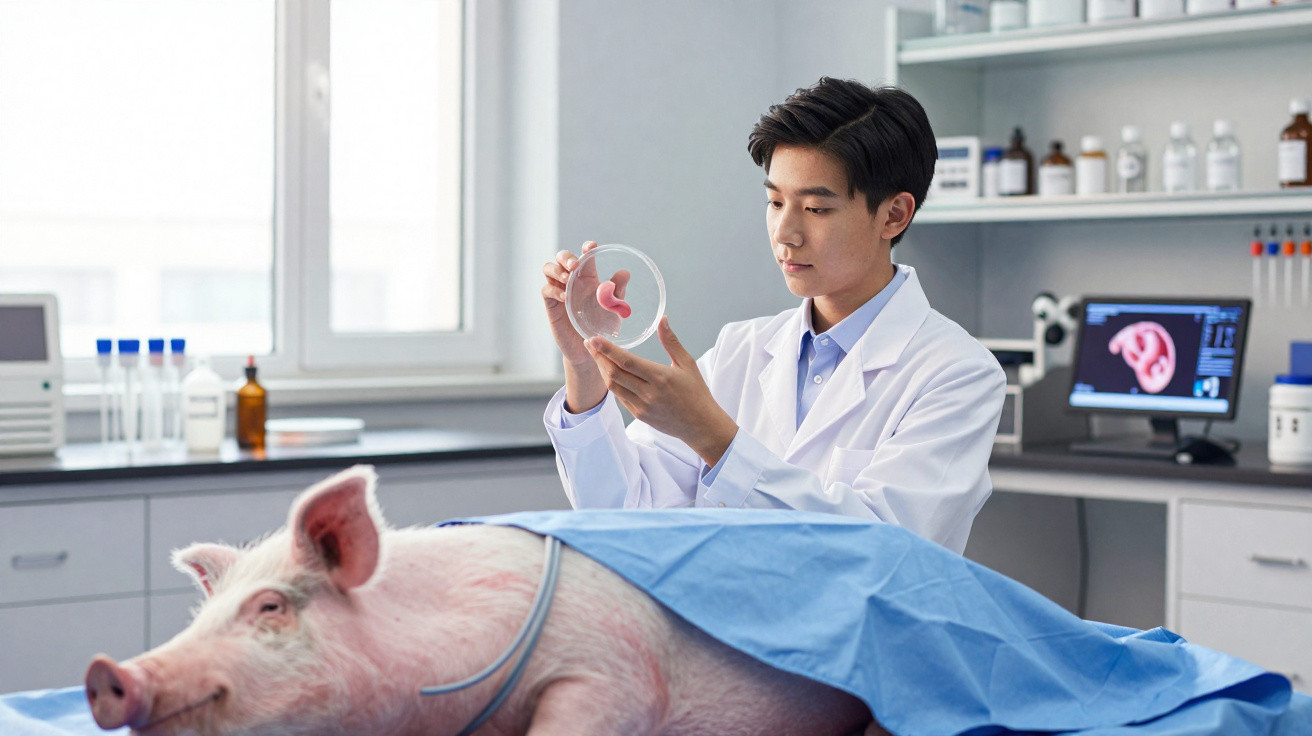
A British research team has, for the first time, successfully grown a section of oesophagus in the laboratory and transplanted it into pigs. The animals were then able to eat normally again. Although it may sound like science fiction, specialists see it as a potential turning point for children with severe malformations and for patients who need part of their oesophagus removed.
What the researchers achieved
The work centres on a segment of the oesophagus: the muscular tube that carries food from the mouth to the stomach. Each of eight miniature pigs received an oesophageal section measuring around 2.5 centimetres, prepared entirely in the laboratory beforehand. The process used the animals’ own cells.
Five of the eight pigs survived the full six-month study period and resumed normal feeding – with a bioengineered oesophagus that behaved like real tissue.
The findings were published in the journal Nature Biotechnology and have attracted international attention because they tackle a long-standing challenge in paediatric surgery: replacing larger sections of the oesophagus.
How a pig organ became a living “scaffold”
The study was led by Paolo De Coppi, a paediatric surgeon at University College London. His team applied a bioengineering technique, beginning with a normal pig oesophagus. It was treated to remove every living cell.
What remained was a framework known as the extracellular matrix. This scaffold:
- retains the organ’s original shape and structure;
- contains no foreign cells that could trigger a rejection response;
- provides a stable foundation for the recipient’s new cells.
The researchers then introduced cells from each individual pig into this biological “skeleton”. These were muscle cells that had been returned to a stem cell-like state. This allowed them to develop into the range of cells required in an oesophagus, including muscle and nerve cells.
The prepared oesophageal section spent one week in a bioreactor. Nutrient solutions circulate within it, while temperature and pressure are kept under controlled conditions. This enabled the cells to attach, multiply and arrange themselves. From the initial organ retrieval to the completed implant took just under two months.
Oesophagus transplantation in miniature pigs: a living-body test
The crucial stage came next. Surgeons removed part of the native oesophagus from eight miniature pigs, each weighing roughly ten kilograms. They filled the resulting gap with the laboratory-prepared segment.
To protect the developing tissue during the first few weeks, the researchers wrapped the implant in a biodegradable mesh. This was intended to encourage the growth of new blood vessels and provide stability until the tissue had integrated.
According to the published data:
- all eight pigs survived the critical first 30 days;
- after three months, the implants had fully integrated in the animals;
- five animals were able to swallow food normally for more than six months.
In these animals, the implant developed:
- muscle capable of contracting;
- nerve structures;
- a functioning vascular network that supplied the segment with blood over the long term.
Measurements showed that the laboratory-grown section of oesophagus could generate sufficient pressure to move food reliably towards the stomach. In some cases, narrowings known as stenoses developed. The team widened these by endoscopy, using an internal tube and balloon – an approach already used to treat people.
Why this offers hope for children with malformations
Children with a congenital malformation called oesophageal atresia are a particular focus. In this condition, the oesophagus has not formed completely. Long-gap defects, in which a substantial section is missing, are especially challenging for surgeons.
Doctors currently often rely on the following stopgap solutions:
- moving part of the stomach into the chest;
- bringing up a section of colon to form a replacement tube;
- complex stretching operations to join the two ends of the existing oesophagus.
Every one of these methods carries risks, ranging from reflux and swallowing difficulties to repeated operations. A personalised implant populated with the patient’s own cells and able to grow with the child could make many of these procedures unnecessary, or at least reduce them substantially.
A scaffold obtained from animal tissue and populated with the patient’s own cells avoids strong immune reactions and adapts to growth – exactly what children with complex malformations need.
Next step: longer oesophageal sections and less manual work
The British group is now working to produce longer segments of between 10 and 15 centimetres. Blood supply is the principal challenge at this stage. The longer the implant, the more difficult it becomes to ensure that every area receives oxygen and nutrients in time.
At the same time, the researchers are standardising the manufacturing process. Their aim is a type of “semi-finished product” made from prepared pig tissue that can be stored in specialist centres. Once a patient has been identified, clinicians would populate this scaffold with that individual’s cells.
This approach could offer several benefits:
- faster availability in emergencies;
- consistent implant quality;
- less manual work in the laboratory;
- avoidance of permanent immunosuppression, because no foreign cells are present.
Study leader De Coppi considers a first clinical trial in humans conceivable in around three to four years, provided the ongoing animal studies involving longer segments produce positive and safe results.
Who else could benefit: cancer patients and caustic-injury survivors
The technique is not intended solely for children with congenital defects. Adults could benefit as well. Following oesophageal cancer, surgeons often remove large parts of the organ. Severe burns caused by cleaning products or alkalis, particularly in household accidents, can be similarly devastating.
At present, doctors generally replace missing sections of oesophagus with parts of the stomach or bowel. This saves lives, but it often results in lifelong limitations in everyday life, from chronic heartburn to swallowing disorders and weight problems. A precisely fitted implant that behaves as much like a natural oesophagus as possible could provide a gentler alternative.
How safe is a bio-implant like this?
Despite the enthusiasm, many questions remain. Could such an implant last for decades? How frequently do narrowings develop? How does the tissue change as a child grows or as a person ages?
The data so far from the pig study provide some initial answers:
| Aspect | Observation in the study |
|---|---|
| Survival after surgery | All eight animals survived the first critical phase |
| Function after 6 months | Five animals ate and swallowed effectively |
| Tissue development | Muscle, nerves and blood vessels were detectable in the implant |
| Complications | Narrowings treated with endoscopic dilation |
Long-term data covering many years are naturally not yet available. Future studies will need to provide these, first in animal models and then in carefully selected patients with very severe underlying conditions.
Technical terms explained
Extracellular matrix
This refers to the scaffold material found between cells. It consists mainly of proteins such as collagen and gives organs their shape and stability. In the current study, this framework is exactly what remains after all living cells have been removed.
Bioreactor
A bioreactor is essentially a “high-tech incubator” for cells and tissues. Temperature, nutrients, oxygen and sometimes pressure or movement can all be set precisely. It therefore creates an environment that imitates the body as closely as possible.
Stem cell-like cells
Although the cells used originally come from muscle tissue, they are reprogrammed in the laboratory so that they can once again develop in different directions. This makes it possible for muscle, nerves and other cell types to form within the implant.
Risks that still remain
Even though the concept appears promising, experts expect a number of obstacles. It remains unclear, for example, whether long-term changes could arise in human implants, such as scarring or abnormal tissue growth. It is also not yet certain how reliably the nervous system functions in such a reconstructed section, as finely coordinated swallowing movements depend heavily on nerve signals.
There are practical issues too: how can these implants be manufactured and funded worldwide? Which hospitals have the expertise to perform the operations safely? And how can it be guaranteed that every individual implant meets the same quality standard?
For all these unresolved questions, the work from London shows that a laboratory-built oesophagus can, in principle, function in larger animals. For families whose children are missing a vital section of this organ, it represents a rare sign of genuine, tangible hope – not in the distant future, but possibly in the years ahead.











Comments
No comments yet. Be the first to comment!
Leave a Comment